Cysts in Venice, FL
What are Cysts?

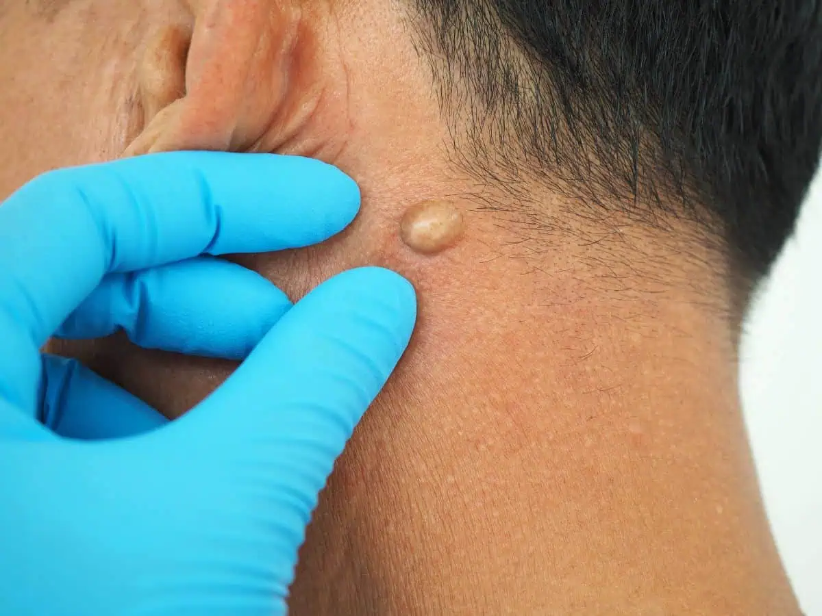

Cysts are small, closed sacs that form under the skin and are filled with fluid, pus, or other material. They can appear anywhere on the body, including the face, neck, back, scalp, or chest. Most cysts are harmless, but they can become uncomfortable, swollen, or infected if left untreated.
Cysts vary in size and type. Some are small and painless, while others may grow larger and cause tenderness. Even though most cysts are noncancerous, professional evaluation is important to confirm the diagnosis and ensure proper treatment.
At REJUVA Dermatology in Venice, FL, our dermatology experts provide safe and effective cyst evaluation and treatment to help relieve discomfort, prevent infection, and improve your skin’s appearance.
What Causes Cysts?
Cysts form when the body’s natural healing or cell renewal process becomes blocked. This blockage causes fluid, oil, or other substances to collect under the skin. Several factors can lead to cyst formation, including:
- Blocked Hair Follicles or Oil Glands: When a follicle or gland becomes clogged, it can form a buildup of oil and dead skin cells.
- Infections: Certain bacterial infections can cause pus-filled cysts to develop.
- Skin Trauma: Injury or irritation to the skin can sometimes trigger cyst formation.
- Genetic Conditions: Some inherited disorders may make people more likely to develop cysts.
- Hormonal Changes: Hormonal shifts can increase oil production and contribute to cyst formation, especially during puberty or pregnancy.
- Foreign Objects Under the Skin: In rare cases, small foreign materials such as splinters can lead to cyst development.
Identifying the type of cyst and what caused it helps determine the best treatment option.
Cysts Symptoms and Signs
- A smooth, round bump under the skin
- Skin that feels firm or slightly soft over the lump
- Swelling or redness if infected
- Pain or tenderness in the area
- Drainage of thick, yellowish fluid if the cyst bursts
- Slow growth over time
Cysts can range from pea-sized to much larger. While many are harmless, it’s important to have them examined, especially if they change in size, color, or become painful.


Treatments for Cysts
- Drainage or Aspiration For cysts that are swollen or uncomfortable, a dermatologist may drain the fluid using a sterile technique. This provides quick relief, although the cyst may return if the capsule remains.
- Surgical Removal Minor surgical removal is often the most effective way to completely eliminate a cyst and prevent it from coming back. This procedure is usually done under local anesthesia in the office and leaves minimal scarring.
- Corticosteroid Injections If the cyst is inflamed but not infected, corticosteroid injections can reduce swelling and discomfort. This treatment helps flatten the cyst without immediate removal.
- Antibiotic Treatment If a cyst becomes infected, oral or topical antibiotics may be prescribed to treat the infection before removal.
- Laser or Electrosurgery Certain types of cysts can be treated with laser or electrosurgical procedures to help remove the cyst wall and reduce recurrence.
After treatment, patients are given care instructions to ensure proper healing and prevent future cyst formation.
Frequently Asked Questions
Are cysts dangerous?
Most cysts are harmless and noncancerous. However, if they become painful, infected, or grow quickly, it’s important to have them checked by a dermatologist.
Can I pop a cyst at home?
No. Popping or squeezing a cyst can lead to infection, scarring, or recurrence. It’s safest to have a dermatologist treat it using sterile methods.
Will a cyst go away on its own?
Some small cysts may shrink over time, but most remain until properly drained or surgically removed. Professional treatment helps prevent infection and recurrence.
Are cysts contagious?
No, cysts are not contagious. They cannot be spread through contact with others.
How can I tell if my cyst is infected?
If the area becomes red, swollen, painful, warm, or begins to drain pus, it may be infected. Seek medical attention right away to prevent further complications.
Will the cyst come back after treatment?
If the cyst wall or capsule is not completely removed, it can grow back. Surgical removal provides the best chance of preventing recurrence.
Can cysts appear anywhere on the body?
Yes. Cysts can develop anywhere, but they are most common on the scalp, back, neck, and face. Some types also form inside the body and require imaging for diagnosis.
Get Relief from Painful or Unwanted Cysts
If you have a cyst that’s uncomfortable, infected, or affecting your confidence, the team at REJUVA Dermatology can help. Our dermatologists provide safe, effective treatments to remove cysts and restore healthy skin.
Schedule your appointment today and let our experts take care of your skin with precision and care.
Experience the best in dermatology care with our expert team of doctors.
Take the first step toward healthier, glowing skin. Our caring dermatology team is ready to help you feel confident in your own skin.
Fill out our quick form or call us today to book your appointment.